Conjoint Tendon Shoulder Anatomy - Anatomy of the RTC tendons - right shoulder. | Download ... / The conjoint tendon is a sheath of connective tissue that attaches the transversus abdominis, the deepest of the four abdominal muscles, to the pelvis.
Conjoint Tendon Shoulder Anatomy - Anatomy of the RTC tendons - right shoulder. | Download ... / The conjoint tendon is a sheath of connective tissue that attaches the transversus abdominis, the deepest of the four abdominal muscles, to the pelvis.. One tendon might have it worse, but it's never isolated to just one tendon. The conjoint tendon (previously known as the inguinal aponeurotic falx) is a structure formed from the lower part of the common aponeurosis of the internal oblique muscle and the transversus abdominis as it inserts into the crest of the pubis and pectineal line immediately behind the superficial inguinal ring. Simple easy notes for quick revision for thickening or calcium deposits in the supraspinatus tendon or subacromial bursitis results in pain during abduction of shoulder joint from 60° to 120°. Normal anatomy and pathology on mri. These are the main ligaments that help to stabilize the joints of.
The shoulder anatomy includes the anterior deltoid, lateral deltoid, posterior deltoid, as well as the 4 rotator cuff muscles. Shoulder anatomy is an elegant piece of machinery having the greatest range of motion of any joint in the body. Related online courses on physioplus. The tendons are the attachment of the. Anterior graphic of the shoulder.
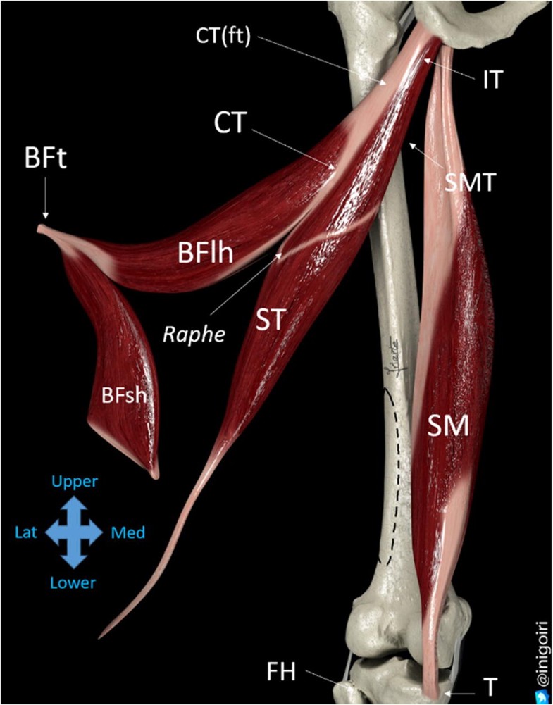
Sonographic landmarks in hamstring muscles | SpringerLink from media.springernature.com If you tear your biceps tendon at the shoulder, you may lose some strength in your arm and have pain when you forcefully turn your arm from palm down to palm up. A muscle contracts to move bones; The shoulder is made up of three bones: Three bones come together at the shoulder joint. Normal anatomy, variants and checklist. The interfoveolar ligament, seen from in front. (inguinal aponeurotic falx labeled at lower left.) falx (disambiguation) — other parts of the anatomy with names including falx. The shoulder is comprised of a ball (humerus) and socket (scapula), bones, ligaments, tendons and muscles that move the arms and connect them to the torso.
Your biceps tendons attach the biceps muscle to bones in your shoulder and in your elbow.
Your biceps tendons attach the biceps muscle to bones in your shoulder and in your elbow. One tendon might have it worse, but it's never isolated to just one tendon. Rotator cuff , a network of muscles and tendons that cover the top of the humerus, or upper arm bone, to hold it place and enable the arm to rotate. (inguinal aponeurotic falx labeled at lower left.) falx (disambiguation) — other parts of the anatomy with names including falx. The shoulder is made up of three bones: The shoulder is comprised of a ball (humerus) and socket (scapula), bones, ligaments, tendons and muscles that move the arms and connect them to the torso. Scapula and related structures — the scapula is a relatively large, flat bone located on the posterior thorax (figure long head of the biceps brachii tendon — the shoulder examination begins by imaging the long head of biceps brachii tendon. An introduction to the anatomy of the shoulder. Normal anatomy, variants and checklist. Shoulder muscles and shoulder tendons. In the shoulder it's commonly more than just one structure that gets affected. Related online courses on physioplus. It reduces wear and tear.
Revision anterior stabilization of the shoulder presents a challenge to the surgeon and carries a higher risk of recurrent dislocation than primary repair. One tendon might have it worse, but it's never isolated to just one tendon. Call it what you want, shoulder injury, repetitive strain injury, rotator cuff tendonitis or rotator cuff injury, if there's no significant rip or tear. Scapula and related structures — the scapula is a relatively large, flat bone located on the posterior thorax (figure long head of the biceps brachii tendon — the shoulder examination begins by imaging the long head of biceps brachii tendon. The biceps muscle has two tendons at the shoulder, called the long head and short head.
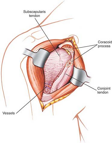
The Shoulder | Musculoskeletal Key from musculoskeletalkey.com Upper limb trauma programme injuries. (inguinal aponeurotic falx labeled at lower left.) falx (disambiguation) — other parts of the anatomy with names including falx. The conjoint tendon can be describe as a layer of connective tissue which connects the pelvis to the transversus abdominis, the deepest of the 4 muscles of the abdomen. Scapula and related structures — the scapula is a relatively large, flat bone located on the posterior thorax (figure long head of the biceps brachii tendon — the shoulder examination begins by imaging the long head of biceps brachii tendon. It gets its name from the fact that it is often continuous or conjoined with the tendon of the internal oblique, another of the abdominal muscles. An introduction to the anatomy of the shoulder. At the shoulder, the two tendons both attach to the large flat bone in the upper trunk called the scapula. The conjoint tendon then turns inferiorly and attaches on.
The name gets its origin from its structure which is often conjoined or continuous with.
It reduces wear and tear. The shoulder joint is the connection between the chest and the upper extremity. It gets its name from the fact that it is often continuous or conjoined with the tendon of the internal oblique, another of the abdominal muscles. The rotator cuff is a group of four muscles and tendons that surround the glenohumeral joint. The conjoint tendon can be describe as a layer of connective tissue which connects the pelvis to the transversus abdominis, the deepest of the 4 muscles of the abdomen. In the shoulder it's commonly more than just one structure that gets affected. Robin smithuis and henk jan van der woude. Shoulder tendonitis is the inflammation, irritation and swelling of the tendons in the rotator cuff and bicep. Upper limb trauma programme injuries. Related online courses on physioplus. Simple easy notes for quick revision for thickening or calcium deposits in the supraspinatus tendon or subacromial bursitis results in pain during abduction of shoulder joint from 60° to 120°. The muscle belly then crosses the entire upper arm and separates into two tendons. Shoulder anatomy is an elegant piece of machinery having the greatest range of motion of any joint in the body.
Robin smithuis and henk jan van der woude. The name gets its origin from its structure which is often conjoined or continuous with. There are a few ways you can help prevent shoulder tendonitis occuring. Professor of radiology and orthopaedic surgery department of radiology university of california san francisco. One tendon might have it worse, but it's never isolated to just one tendon.
Anatomy Of Shoulder Bones Ideas Shoulder Anatomy Medical ... from i.pinimg.com These are the main ligaments that help to stabilize the joints of. At the shoulder, the two tendons both attach to the large flat bone in the upper trunk called the scapula. The conjoint tendon can be describe as a layer of connective tissue which connects the pelvis to the transversus abdominis, the deepest of the 4 muscles of the abdomen. Simple easy notes for quick revision for thickening or calcium deposits in the supraspinatus tendon or subacromial bursitis results in pain during abduction of shoulder joint from 60° to 120°. The interfoveolar ligament, seen from in front. Normal anatomy, variants and checklist. Revision anterior stabilization of the shoulder presents a challenge to the surgeon and carries a higher risk of recurrent dislocation than primary repair. The conjoint tendon then turns inferiorly and attaches on.
Shoulder tendonitis is the inflammation, irritation and swelling of the tendons in the rotator cuff and bicep.
Three bones come together at the shoulder joint. Learn vocabulary, terms and more with flashcards, games and other study tools. Shoulder muscles and shoulder tendons. One tendons inserts onto the forearm bone, the radius, and the second spreads out to join the fascia. There are several important ligaments in the shoulder. Your biceps tendons attach the biceps muscle to bones in your shoulder and in your elbow. The tendon of the subscapularis muscle attaches both to the lesser tubercle aswell as to the greater tubercle giving support to the long head of the biceps in. Scapula and related structures — the scapula is a relatively large, flat bone located on the posterior thorax (figure long head of the biceps brachii tendon — the shoulder examination begins by imaging the long head of biceps brachii tendon. First, you need to warm up and stretch the shoulder area before any sport or activity that is going to place strain on. The muscle belly then crosses the entire upper arm and separates into two tendons. Revision anterior stabilization of the shoulder presents a challenge to the surgeon and carries a higher risk of recurrent dislocation than primary repair. Professor of radiology and orthopaedic surgery department of radiology university of california san francisco. If you tear your biceps tendon at the shoulder, you may lose some strength in your arm and have pain when you forcefully turn your arm from palm down to palm up.
Three bones come together at the shoulder joint shoulder tendon anatomy. Professor of radiology and orthopaedic surgery department of radiology university of california san francisco.